Category Archives: Biological Illustration

A pencil drawing for a change of pace and a better world

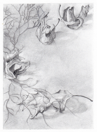
Every year, Mr. Saad Ghosn publishes a book entitled “For a Better World, Book of Poems and Drawings on Peace and Justice by Greater Cincinnati Artists.”  He asks the artists what kind of poetry they like, and then pairs them up with a few different poems to illustrate.  This year the book is in its eighth edition, and I am excited to be contributing this drawing…

Pencil and charcoal on paper

It goes with a poem that uses trees as a metaphor to liken dreams, family, and memories to the life/growth/death/new life cycle (at least, that was my interpretation) by Kathryn Martin Ossege. It’s been a while since I did a nice complete drawing without any software component, and I forgot how relaxing it was.  I look forward to the book coming out and getting to see all of the other drawings and poems that were contributed.

Live cell 3D microscopy

Accuracy is important to medical illustrators’ work.  But what if you cannot see the subject matter you are drawing?  I love illustrating the world of the cell, so I am always on the look-out for new resources and references that can show me what cells and intercellular structures actually look like.  I recently heard about a new microscope that can create 3D movies of live cells.  Located at the Howard Hughes Medical Institute, this microscope uses what is known as a Bessel beam, a band of light which scans rapidly through live cells, capturing images (200 per second) that can be stacked together to show the 3D structure.  See the full article at http://www.hhmi.org/news/betzig20110304.html.

Because this technique does not require the cells to be fixed and stained, nor does it kill them by over-exposing them to too much light, their structure can be examined over time, showing their membranes and organelles moving.

For example, one such movie shows the chromosomes separating over the course of 18 minutes, in preparation for cell division (still image shown below).

Mitosis (screen shot from video at http://www.hhmi.org/news/popups/mitosis_mov_pop.html)

This research was carried out at HHMI’s Janelia Farm Research Campus, where one of their research focuses is the development of imaging technologies and computational methods for image analysis.  Some rainy day, I’ll have to browse their archives and catch up on the latest developments in microscopy.

Virus

Created in Adobe Illustrator

It’s that time of year, and it seems there is a lot of nasty stuff going around right now, so I am posting this virus doodle I did in a design class in grad school.  It’s heavily stylized, yet sinister and kind of cute all at the same time.

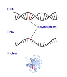
from DNA to protein

DNA (transcription) RNA (translation) Protein!

Created in Illustrator, with the exception of the protein, which was exported from a 3D molecular viewer (JMol).

DNA

Technically, I didn’t draw this.  I used a 3D molecular rendering program to visualize data from the Protein Data Bank.  Access to this data and the program I used, Chimera, is free.

A double strand of DNA